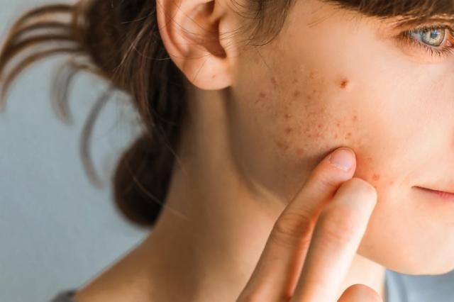
Skin cancer on face
Skin cancer which is the abnormal growth of skin cells — most often develops on sun exposed areas, such as face. But skin cancer can also occur on areas of the body not ordinarily exposed to sunlight.
There are three major types of skin cancer — basal cell carcinoma (BCC), squamous cell carcinoma (SCC) and malignant melanoma (MM).
What is basal cell carcinoma?
Basal cell carcinoma (BCC) is the most common, locally invasive, keratinocytic non-melanoma skin cancer. It is also known as rodent ulcer. It rarely spreads to other parts of the body or kills but it can cause significant destruction and disfigurement by invading surrounding tissues.
It presents as sores, red patches, scaly patches, pink growths, shiny bumps, or scars and is usually caused by a combination of cumulative and intense, occasional sun exposure. BCC almost never spreads (metastasizes) beyond the original tumour site
What is squamous cell carcinoma?
Cutaneous squamous cell carcinoma (cSCC) which is often called squamous cell carcinoma (SCC) is the second most common type of non-melanoma skin cancer. It is derived from cells within the epidermis that make keratin — the horny protein that makes up skin, hair and nails. It can often appear as a firm pink lump with a rough or crusted surface.
It is more common on sun exposed areas such as the head, ears, neck and back of the hands. It can metastasize elsewhere and must be treated early
What is malignant melanoma?
Melanoma is a malignant tumour of melanocytes. Melanocytes are cells that produce the dark pigment, melanin, which is responsible for the color of skin. Melanoma is described as:
- In situ, if the tumouris confined to the epidermis
- Invasive, if the tumourhas spread into the dermis
- Metastatic, if the tumourhas spread to other tissues
What are the risk factors for BCC and SCC?
The main risks factors for developing BCC and SCC are:
- Long term sun exposure
- Older age
- Gender-more common in male patients
- Fair skin, blue or green eyes, blond or red hair
- Previous skin cancer
- Sun damage-actinic keratosis
- Previous cutaneous injury-thermal burn, lupus, sebaceous naevus
- Immunosuppression
- Chronic inflammation
- Ionising radiation and exposure to arsenic
- Family history
What are the risk factors for malignant melanoma?
The main risks factors for developing MM are:
- Cumulative sun exposure
- Older age
- Previous invasive MM
- Previous non-melanoma skin cancer (BCC, SCC)
- Many melanocytic naevi (moles)
- Multiple (>5) atypical naevi
- Strong family history of melanoma with 2 or more first-degree relatives affected
- Tanning beds
- Genetic risk factors-BRAF and p53 mutation
Who is more likely to develop BCC and SCC?
The following groups of people are at greater risk of developing the BCC:
- Immunosuppressed patients
- eople who have had significant long term and cumulative ultraviolet light exposure
- People susceptible to sunburn
- People who use sun beds regularly (need to avoid tanning beds)
- People with inherited syndromes-Gorlin syndrome, xeroderma pigmentosum, Bazex-Dupre-Christol syndrome
Who is more likely to develop MM?
The following groups of people are at greater risk of developing the MM:
- Immunosuppressed patients
- rgan transplant patients
- People who have had significant cumulative ultraviolet light exposure
- People susceptible to sunburn
- Family history of MM
- Familial Atypical Multiple Mole Melanoma Syndrome
- People with many (more than 50) ordinary moles
What are the clinical features of BCC?
BCC can vary in their appearance, but most usually it appears as:
- Slowly growing plaque or nodule. Sometimes the patch crusts
- May be skin coloured, pink or pigmented
- Pink growth with elevated rolled border, crusted indentation in the centre and tiny blood vessels on the surface
- Can bleed and ooze
- Can present as a scar-like area
- May ulcerate
- Vary in size from few millimetres to several centimetres in diameter
- In addition, any change in a preexisting skin growth, such as an open sore that fails to heal, or the development of a new growth, should prompt an immediate review
What are the clinical features of cSCC?
SCC can vary in their appearance, but most usually it appears as:
- Scaly or crusty raised area of skin with a red, inflamed base
- Hard plaque or a papule
- May look like warts
- May be painful or tender
- Can bleed or may ulcerate
- Grows over weeks or months
- Vary in size from few millimetres to several centimetres in diameter
- In addition, any change in a pre-existing skin growths, such as an open sore that fails to heal, or the development of a new growth, should prompt an immediate review
What are the clinical features of MM?
MM can vary in the appearance, but most usually it appears as:
- Unusual looking freckle or mole
- Variety of colours, including no pigment
- Can be flat or raised
- Can be itchy or tender
- New mole or an existing one that is growing, changing colour (either becoming lighter or darker) or becoming irregular in some way
How is BCC and BCC diagnosed?
Diagnosis of skin cancer is based on clinical features. To confirm the diagnosis, a small piece of the abnormal skin (a biopsy), or the whole area (an excision biopsy), is removed under a local anaesthetic and sent to a pathologist to be examined.
Some typical superficial BCCs on trunk and limbs are clinically diagnosed and have non-surgical treatment without biopsy.
Patients with high-risk SCC may also undergo staging investigations to determine whether it has spread to lymph nodes or elsewhere.
How is MM diagnosed?
- History
- Examination
- ABCDE
- Dermoscopy
- Biopsy
- Dependent on size of lesion and patient
- Excisional biopsy, 2mm margin
- Patient counselling
- Imaging maybe considered
- SLNB maybe considered
How can BCC and SCC be treated?
If caught early, they are curable and cause minimal damage. However, the larger and deeper a tumor grows, the more dangerous and potentially disfiguring it may become, and the more extensive the treatment must be. The treatment used will depend on the type, depth of penetration, size and location of the skin cancer, as well as the patient’s age and general health.
Most skin cancers are treated surgically. This involves removing the BCC or SCC with a margin of normal skin around it (3-4mm), using a local anaesthetic. The skin is then closed with stitches or defect is reconstructed with a local flap or skin graft.
Sometimes other surgical methods are used such as Mohs surgery, curettage, cryotherapy, topical anti-cancer ointments, radiotherapy, combination of treatments for advanced skin cancers.
How can MM be treated?
Three quarters of the people who have a melanoma removed will have no further problems. The thinner the melanoma is when it is removed; the better is the survival rate. In a small minority of people, the melanoma may have spread but further surgery or immune chemotherapy can often help to control this.
Surgery is usually the recommended treatment. This involves removing the suspicious mole with a 2mm margin of normal skin around it, using a local anaesthetic. The skin is then closed Once the Breslow thickness is determined a wide local excision is usually undertaken. The width of excision size is determined by the depth and stage of the melanoma. It is important this is performed by a team experienced in melanoma excision that is part of the skin cancer Multi-Disciplinary Team (MDT) team.
Dependent on stage, a patient may also be offered a Sentinel Lymph Node Biopsy (SLNB) to check if the cancer has spread to local nodes. This is carried out under general anaesthetic at the same time as the wider excision. A positive result means that cancer has started to spread. In New Zealand, many surgeons recommend sentinel lymph node biopsy for melanomas thicker than 1 mm, especially in younger persons. However, although the biopsy may help in staging the cancer, it does not offer any survival advantage. The necessity for sentinel node biopsy is controversial at present.
When to see a doctor
Make an appointment with your doctor if you notice any changes to your skin that worry you. It is important to examine your skin. Not all skin changes are caused by skin cancer. Your doctor will investigate your skin changes to determine a cause.

Recent Comments